Hydatid Cyst Of Liver And Other Abdominal Organs
TAn infection with the larval stage of dog tapeworm or hydatid worm Echinococcus granulosus causes hydatid disease in humans. Occurring due to the ingestion of tapeworm eggs excreted in the faeces of infected dogs, it is an important pathogenic, parasitic, and zoonotic infection of humans. In certain parts of the world, hydatid disease is a major endemic health issue.
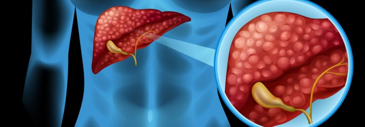
Cystic hydatid disease most often affects the liver (50–70%), but it may also affect other abdominal organs. After a cyst ruptures into the biliary tract or peritoneum, liver hydatidosis may cause dissemination or anaphylaxis. Infection of the cyst can lead to liver abscesses and local complications.
Complications of Hydatid Cysts
In about 40% of the cases, echinococcal cysts in the liver may cause complications. Infection, biliary tree rupture, peritoneal cavity rupture, and pleural cavity rupture are the most common complications, in order of frequency, says the liver doctor in Kolkata. However, GI tract, bladder, and vessel ruptures are extremely uncommon.
Diagnosis of Hydatid Cysts
Humans are normally asymptomatic for a long time after contracting echinococcal infections. The size of the cyst in the liver varies from year to year, varying from 1 mm to 5 mm in diameter. The majority of primary infections have only one cyst, but up to 20% – 40% of infected people have multiple cysts.
Non-Complicated Cysts
The presence of a hydatid cyst in the liver is often overlooked, and it is only discovered by chance during an abdominal examination for another condition. The clinical symptoms emerge progressively as the cyst volume grows larger. When it happens, the most common symptom is right upper quadrant or epigastric pain, with a swollen liver and a palpable mass being the most common test findings.
Complicated Cysts
Patients can also experience complications, including biliary contact, intraperitoneal rupture (spontaneous or post-traumatic), and, in extreme cases, intrathoracic or intrapericardial rupture. The liver doctor says that the cyst rupture can cause anaphylaxis due to the cyst fluid’s high antigenic content, or it can be quiet and present with multiple intraperitoneal cysts.
Ultrasound or other imaging methods such as CT-scan or MRI, in combination with case history, are the easiest ways to make a diagnosis. In addition, serology tests like ELISA or immunoblotting can be used, which are 80-100 percent sensitive for liver cysts but just 50-56 percent sensitive for other abdominal organs.
Hydatid Cyst Treatment
The gold standard treatment for hydatid cyst in liver is surgery. The aim of surgery is to inactivate the parasite, evacuate the cyst with resection of the germinal layer, prevent peritoneal scolices spillage, and destroy the residual cavity. According to the liver surgeon in Kolkata, if a cyst does not have a dangerous location, surgery can be performed successfully in up to 90% of patients.
Surgery, on the other hand, could be impractical in patients with multiple cysts in multiple organs. Chemotherapy and the PAIR technique (puncture-aspiration-injection-respiration) have been introduced as an alternative treatment option, especially for inoperable patients and cases with a high surgical risk.
Take the first step towards a healthier you